Stitching of 3×3 FoVs

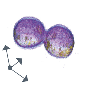
Cell population
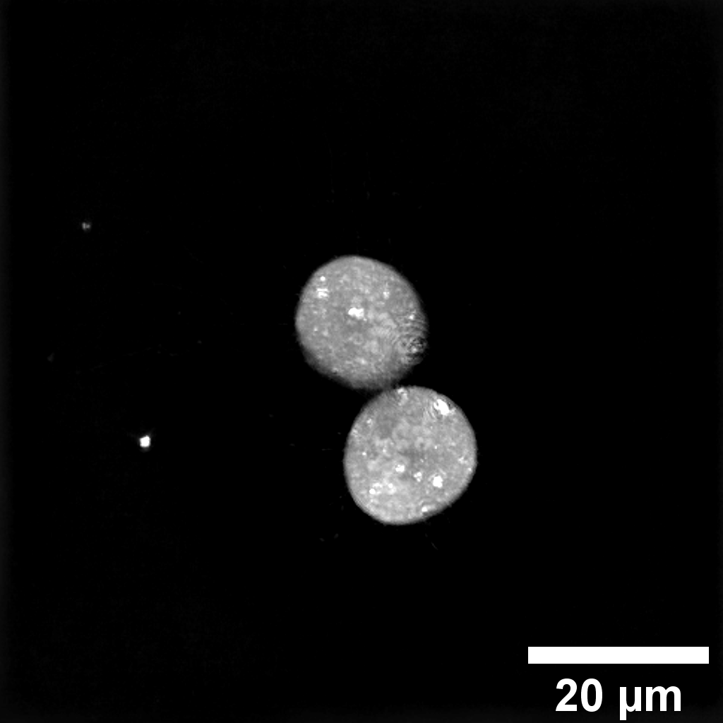
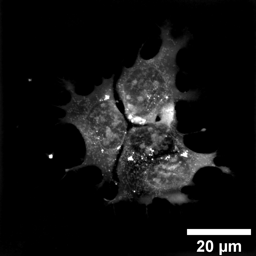
Single FoV

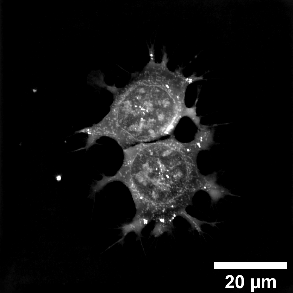
Single cell
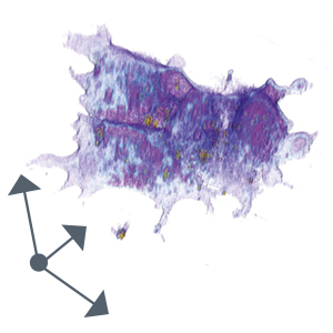
Zoom into FoV

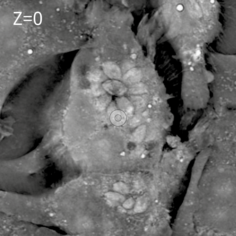
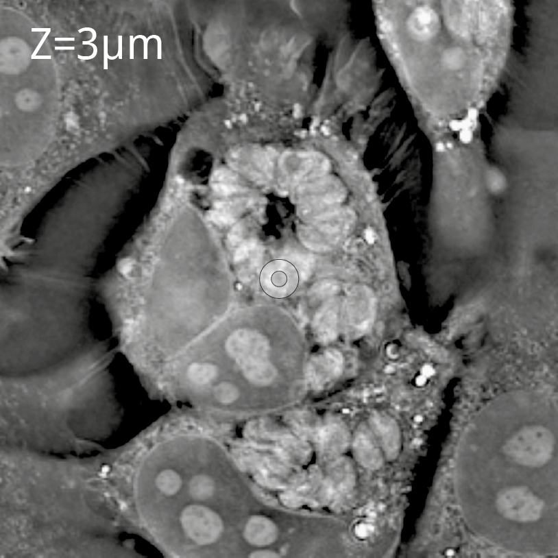
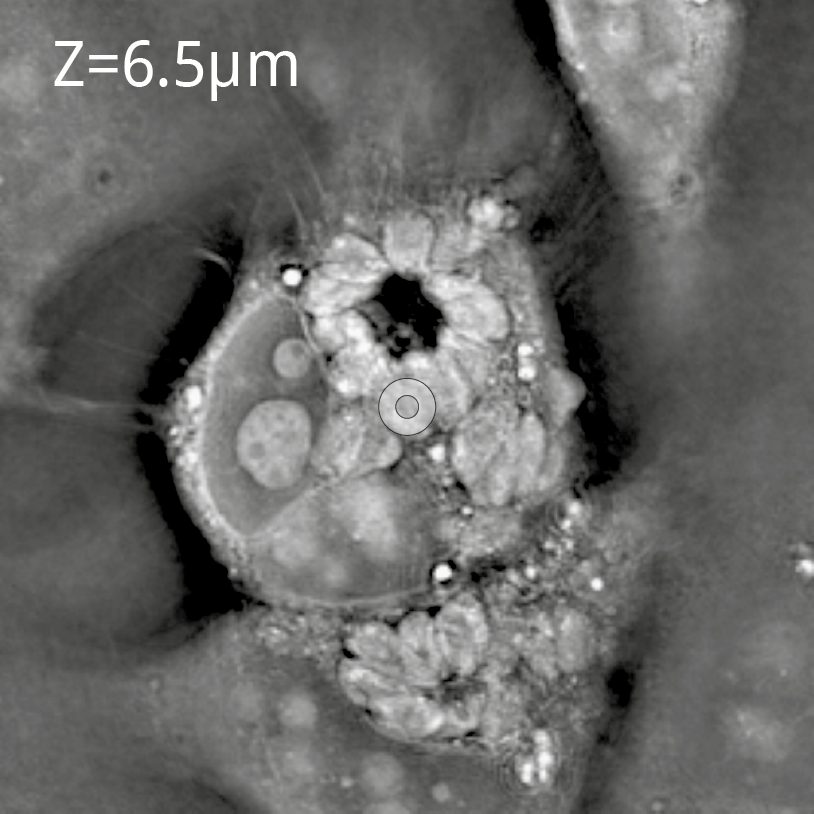
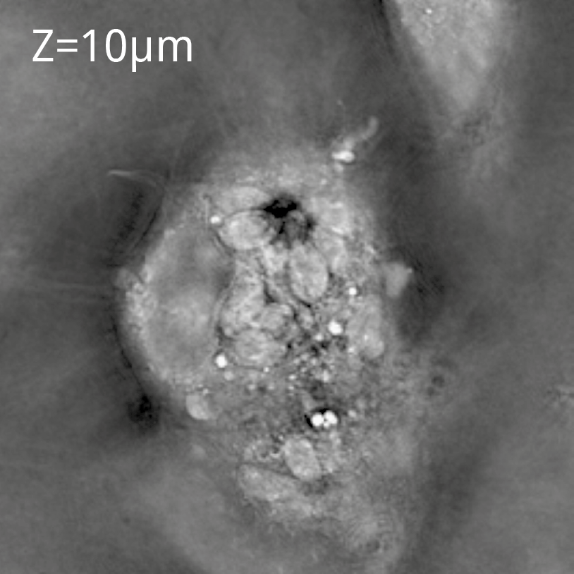
Organelle ecosystem
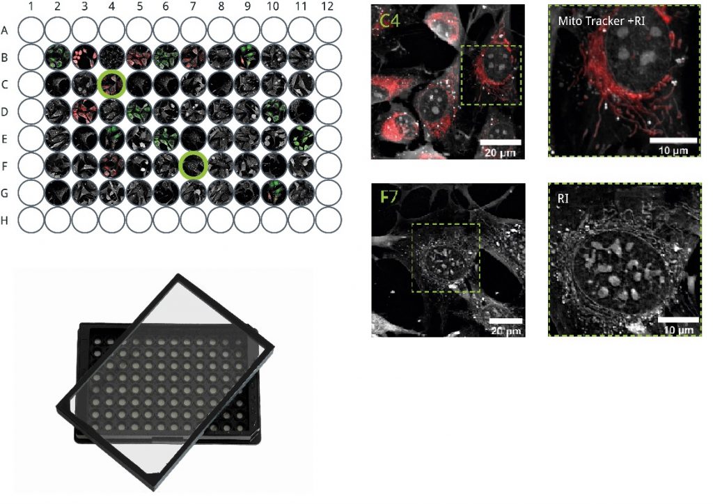

Stitching of 3×3 FoVs
Cell population
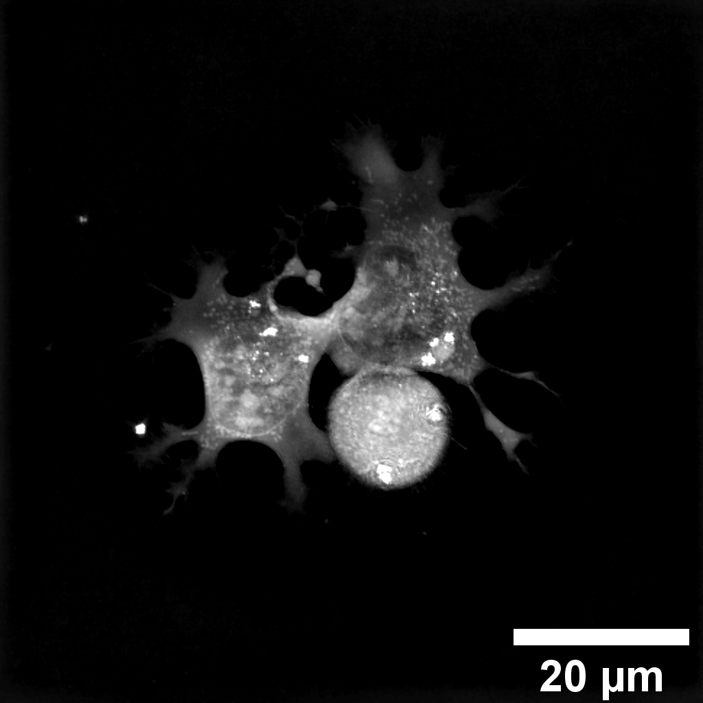
Single FoV
Single cell
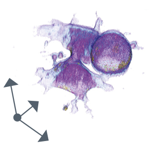
Zoom into FoV
Organelle ecosystem