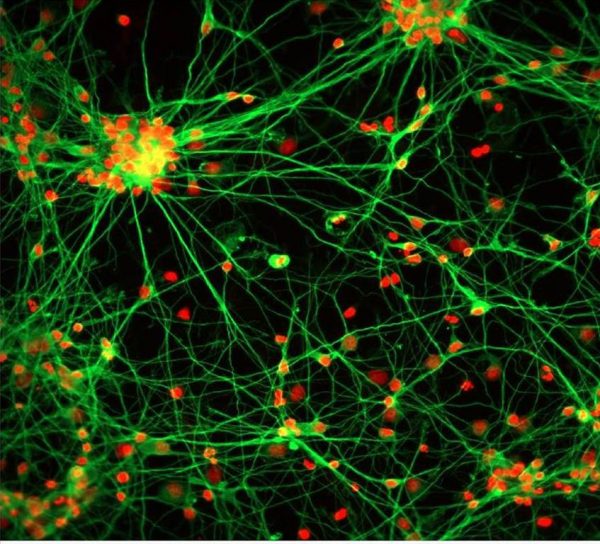
DeltaVision OMX Flex super-resolution microscope
DeltaVision OMX Flex is a multi-imaging mode, super-resolution microscope optimised for structured illumination microscopy (SIM).
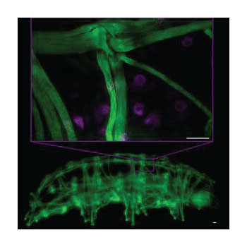
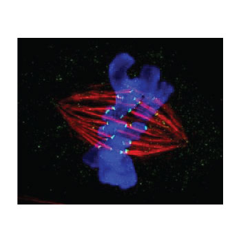

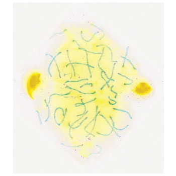
DeltaVision OMX Flex super-resolution microscope
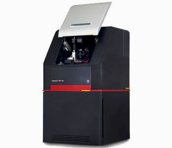
DeltaVision OMX Flex super-resolution microscope accelerates time to publish by revealing insights derived from each of the imaging modes: 3D-, 2D-, TIRF-SIM, ring TIRF, widefield with deconvolution, localization, photokinetics, and EDGE confocal.
- Switch between imaging modes with a click of a button—no hardware alignment needed or changing/moving biological sample.
- Image as fast as your biology requires without having to sacrifice cell image analysis quality or speed.
- Experience two-fold resolution improvement in all three dimensions for an eight-fold volumetric improvement in resolution (3D-SIM).
- Efficient delivery and detection light maximizes contrast, which allows gathering of more data, longer imaging, and minimal photobleaching.
- TIRF-SIM delivers the ability to observe some of the most challenging biological processes in super-resolution at 20 reconstructed fps.
- Simultaneously acquire up to four wavelengths at a time with ultrafast widefield imaging (> 375 fps at 512 × 512).
See a short video comparing widefield with 3D-SIM for individual mitochondria (green) interacting with the endoplasmic reticulum (mCherry) network (Vaughan Lab, University of Washington, 2-color imaging for 200 time points at 96 fps).
Overview
How small are the tissues, cells, and organelles you are studying?
What level of detail do you need to answer your biological questions and achieve your research goals? DeltaVision OMX Flex microscope allows you to use multiple imaging modes from confocal to single molecule localization to go from a macro- to a nanoview of tissues, cells, and organelles.
- Conventional resolution with widefield deconvolution and line-scanning confocal.
- Fast 3D-SIM for volumetric observation at 100 nm resolution.
- DeltaVision localization microscopy for 30 nm level of detail.
How deep beneath the cell membrane layer?
Some biological processes occur at the surface of the cell, while other interactions are deep beneath the cell membrane layer. If you need to go deeper, DeltaVision OMX Flex gives you the power to observe incredibly close to the coverslip in multiple imaging modalities or to build images in 3D of deep samples.
- TIRF microscopy in conventional and SIM modes for exquisite detail close to the coverslip.
- 3D-SIM and deconvolution microscopy for increased resolution and contrast to 50 µm depth.
- Line scanning confocal microscopy for large tissue and cleared samples.
How fast does your imaging speed need to be?
Would faster imaging speeds help you understanding your biological system better? DeltaVision OMX Flex provides fast imaging for the observation of dynamic events whilst also providing an efficient platform for imaging multiple field of views very rapidly.
- Fast reproducible stage with point visiting capabilities.
- 20 TIRF-SIM fps imaging speeds.
- Multiple sCMOS cameras and computers for four color simultaneous acquisition at > 375 fps burst mode.
How many cells are you imaging?
Are you studying events in a single cell or handful of cells? DeltaVision OMX Flex has multiple tools to help you increase the efficiency and volume of your imaging.
- Rapid imaging speeds in all modalities to allow more acquisitions from a single sample.
- Point visiting for a single timepoint or multiple timepoints.
How alive do your cells need to be for cell imaging?
Do you struggle with photobleaching limiting your live cell experiments? Live cell imaging still faces many challenges such as preserving cell health while imaging for extended time periods as well as improving spatial and temporal resolution. DeltaVision OMX Flex microscope was redesigned to focus on reducing background noise, enhancing contrast, and ensuring signal quantitated to deliver high-quality images.
- Image as fast as your biology requires without having to sacrifice speed or image quality with minimal photobleaching.
- Precise control of sample temperature, gas concentration and humidified air to the sample to preserve cell viability.
- Cells remain in a metabolic state with no nonspecific changes that could alter the process being observed.
See a short video of cells undergoing mitosis (Heidi Hehnly, Scott Lab University of Washington & HHMI).
PRODUCT SPECIFICATIONS
DeltaVision OMX Flex – Product Specifications
| Parameter | DeltaVision OMX Flex |
|---|---|
| Dimensions | Door closed: 86 cm (33.7 in.) × 160 cm (62 in.) 92 cm (36.2 in.) Workstation: 43.2 cm (17 in.) × 45.8 cm (18 in.) × 17. 8 cm (7 in.) |
| Camera | sCMOS detector; 2040 × 2040 imaging array; 6.5 × 6.5 μm pixels 16-bit dynamic range; 272 MHz, 95 MHz readout speed options 0.9 (median)/1.4 (rms) e-readout noise |
| Illumination | Structured illumination: 3D-SIM, 2D-SIM, total internal reflection fluorescence (TIRF)-SIM; IRIS/EDGE line-scanning confocal; widefield with deconvolution; ring TIRF; photokinetics/photoactivation (PK/PA); localization microscopy (DLM); brightfield illumination and differential interference contrast (DIC) |
| Standard supported dyes and fluorophores | Blue (DAPI, Hoechst, CF™405M) Green (GFP, Cy2, Alexa Fluor™ 488, ATTO-488, CellTracker™ Green, Calcein AM) Red (mCherry, mKate2, Alexa Fluor 568) Far Red (Cy5, Alexa Fluor 647, To-Pro™-3, SiR) |
| Excitation lasers (nm) | 405, 488, 561, 640 |
| Power consumption | 900 VA |
| Humidity | Stable under 50%, noncondensing |
| Stage travel | 25 × 50 × 25 mm |
| Standard objective lens | Certified 60× 1.42 NA PlanApoN PSF |
| Standard supported sample types | Microscope slides (75 × 25 mm); 35 mm dishes; 2, 4, or 8-well chambered coverglass (24 × 60 mm); 2, 4, or 8-well chambered microscope slides (75 × 25 mm) |
| Workstation | CentOS 6 or higher; 32 GB 1600 MHz DDR3; 256 GB SSD OS hard disk; 3 × 1 TB Onboard RAID5 Array |

