CV1000 - All-in-One Confocal Imaging Solution
The Ideal Tool for Long-Term Live Cell Imaging
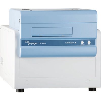
The CellVoyager CV1000 Confocal Scanner System is a fully integrated desktop imaging system.
With its microlens enhanced dual Nipkow disc scanning technology, phototoxicity and photobleaching are drastically reduced, making it ideal for use in observing highly delicate life processes such as iPS/ES cell generation and embryogenesis. The system is easy to use and eliminates the need for a dark room.
Overview
Cell-friendly confocal microscope
- Microlens-enhanced Dual Nipkow Scanning means low phototoxicity / photo bleaching
- Offers clear-cut real confocal images
Most stable environment control
- No need for severe temperature control of your lab
- Capable of really long-term live cell imaging in a dish, covered glass chamber or microplate
You no longer lose the best shot
- Map View function enables quick search for the target area
- Can seamlessly check the full events in life by multi-point time lapse imaging
- Autofocus searches for the cell adhesion plane all the time
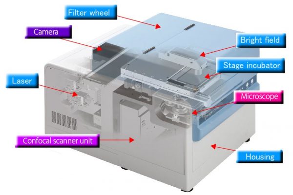
Provides all functions necessary for live cell imaging within a box
- No need for a dark room
- No need for a vibration isolator
Attachment
From high-end multi-point, long-term time lapse imaging to single shots of fixed cells.
Laser Filter
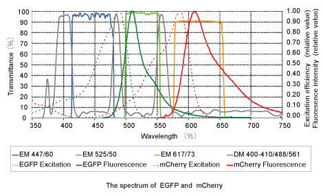
Selectable for up to three laser lines. Selectable from 405nm, 488nm and 561nm, depending on the application. Steep optical separation by the specially designed dichroic mirror in combination with the high-yield/high blocking EM filters enables high-S/N sharp imaging. Installable up to 6 EM filters.
Autofocus
CV1000 autofocus optically detects the glass surface to set it as the Z- offset position. It is useful for not only searching for the objects but useful to avoid focus drift caused by the deflection of glass bottom surface of a dish or a microplate during long-term time lapse. Offers high level of positional repeatability when you observe the same wells in a microplate many times, such as for live cell screening.
Excheangable Pinhole
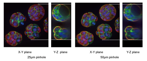
You can select the pinhole size that works best with you chosen magnification, for optimal imaging.
SPECIFICATIONS
| CONFOCAL SCANNER SYSTEM | |||||||
|---|---|---|---|---|---|---|---|
| Model | CV1000 | ||||||
| Main unit | Type | 3-color model | 2-color model | Single-color model | Basic model | ||
| Confocal scanning method | Microlens enhanced dual Nipkow disk scanning | ||||||
| Excitation laser wavelength | 405、488、561 | 488、561 | 488 | 488 | |||
| Bright field imaging | LED transmission | – | |||||
| Camera | Type | Ultra high sensitive Back-illuminated EMCCD | Ultra high resolution Cooled CCD | ||||
| Effective no. of pixels | 512×512 | 1344 ×1024 | |||||
| XY-stage | High-precision auto X-Y stage Designated resolution: 0.1um | ||||||
| Z-axis control | Motorized Z-axis control Designated resolution: 0.1um | ||||||
| Objective lens | [Standard] Dry: 10x [Option] Up to 5 lenses can be added Dry: 20x, 40x Oil: 20x, 40x, 60x, 100x Water: 60x LWD: 20x, 40x Silicon: 30x, 60x | ||||||
| Stage incubator*1 | High-precision temperature controllable incubator Temperature Range: 30 up to 40oC (Room temperature +5o or higher) Designated resolution : 0.1oC Humidity control: Forced humidification with a water bath unit | ||||||
| CO2 | CO2 consentration:1-9% or OFF Gas cylinder*2:CO2 gas | – | |||||
| External dimension(mm) | W580 x D835 x H532 | ||||||
| Weight(kg) | 93 | ||||||
| Utility box | External dimension(mm) | W319 x D368 x H518 | W319 x D368 x H346 | ||||
| Weight(kg) | 16 | 10 | |||||
| Control software | Sets conditions for imaging, camera, time lapse, environments*1, 3D imaging,map view acquisition, multi-color imaging , and multi-point imaging. Functions include image display. Output image file type : 16bit TIFF, JPEG, PNG Output movie file type : AVI | ||||||
| Work station | Controller work station, Display | ||||||
| Operating temperature | 15 up to 35oC (When operating temperature is over 30oC, water cooling of the camera is required.) | ||||||
| Operating humidity level | 20 up to 70% RH (no condensation) | ||||||
| Power consumption | 100-240VAC 1.5KVAmax | ||||||
| OPTION | ||||
|---|---|---|---|---|
| Pinhole change unit | 50um/25um Switching time : 2sec | |||
| Camera | Model | Type | Effective no. of pixels | Pixel size |
| Ultra high sensitive | Back-illuminated EMCCD | 512×512 | 16um | |
| High resolution and sensitive | 1024×1024 | 13um | ||
| Ultra high resolution | Cooled CCD | 1344×1024 | 6.45um | |
| Auto focus | Detection of glass surface with laser + offset | |||
| Attachment | For Single 35mm dish with Stage incubator*1 For Triple 35mm dishes with Stage incubator For Cover glass chamber with Stage incubator For Microplate with Stage incubator For Slide glass *3 For Microplate | |||
| SAMPLE | |||
|---|---|---|---|
| 35mm dish | Vender | Model number | Diameter of glass |
| MatTek | P35G-0-14-C | φ14 | |
| P35G-0-10-C | φ10 | ||
| IWAKI | 3911-35 | φ12 | |
| Matsunami | D110300 | φ14 | |
| Greiner | 627860 | φ20 | |
| ibidi | 81158 | φ21 | |
| 81156 | |||
| BD | 351008 | φ20 | |
| Cover glass chamber | Vender | Model number | Number of wells |
| IWAKI | 5232-008 | 8 | |
| NUNC | 155411 | 8 | |
| 155409 | 8 | ||
| Microplate | Vender | Model number | Number of wells |
| IWAKI | 5816-006 | 6 | |
| 5826-024 | 24 | ||
| 5866-096 | 96 | ||
| 5883-384 | 384 | ||
| Greiner Bio-One | #655896 | 96 | |
| #781896 | 384 | ||
| AURORA | 00019299B.200.EB.ULB | 384 | |
| PerkinElmer | #6007430 | 384 | |
*1 Option (Basic model)
*2 CO2 gas cylinder not included with CV1000 system
*3 Option (3-color model,2-color model and single-color model).


