Expertise in scientific imaging

NANOLIVE

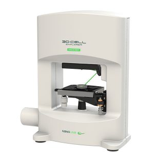
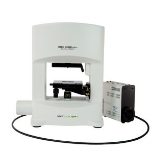
Our 3D Cell Explorer delivers living cell tomography. Just as you may know from a MRI in hospitals, our light microscope uses laser light to deliver live scans of single cells. After placing the cellular specimen in the device, the 3D Cell Explorer performs a continuous rotational scan, while a software allows to display the cell on your computer in 3D within a second.

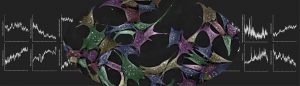
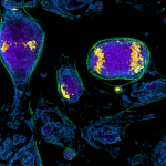
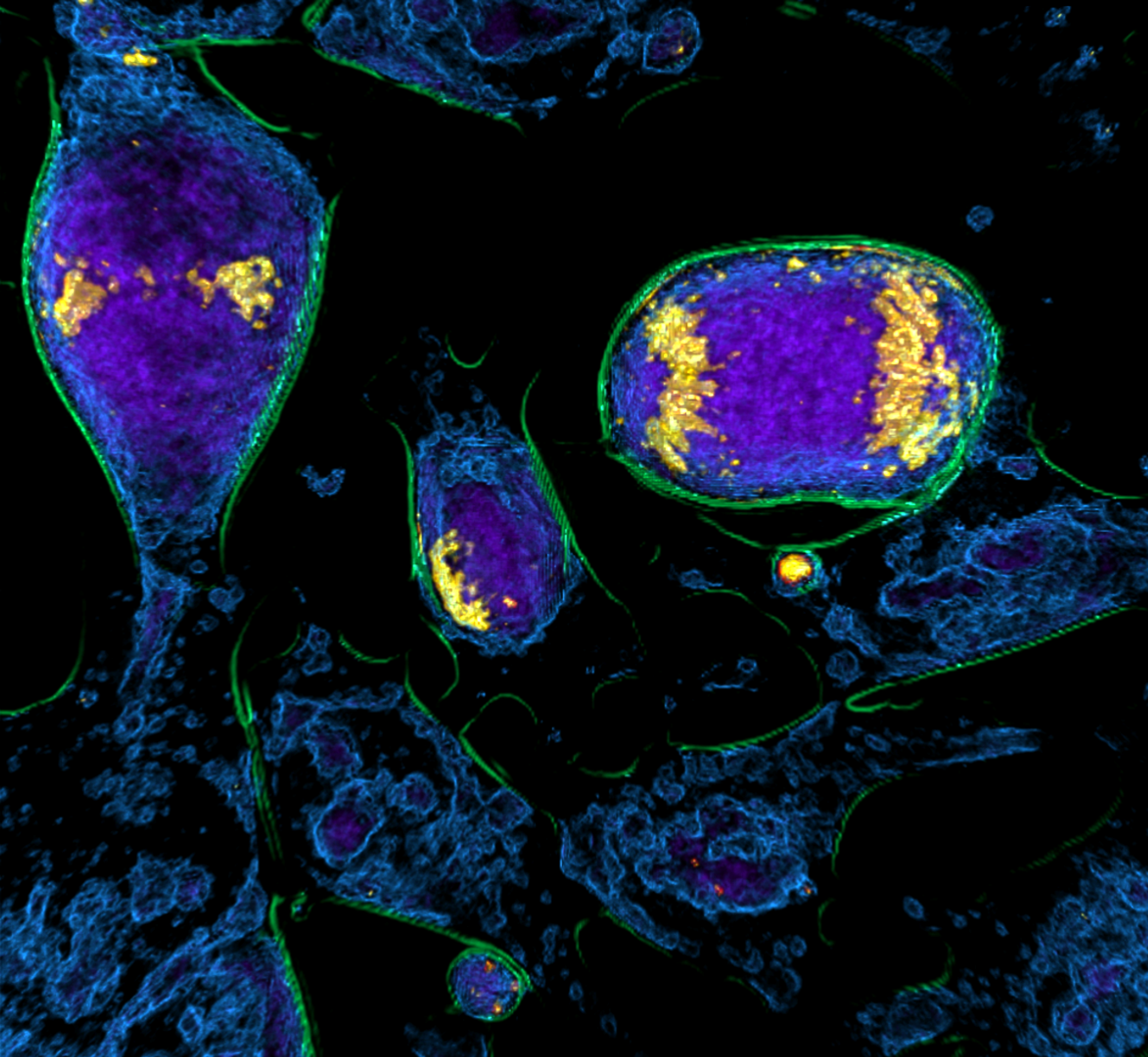
The intuitive software STEVE enables digitally staining on single cells with an unlimited choice of colours and obtains its 3D reconstruction in real time.

To share, interact, and explore your results, the cells data can further be printed (e.g. 3D printer or 3D holograms), or can be directly viewed on 3D-beamers or in 3D animations.

The 3D Microscopes

A revolutionary Tomographic Microscope to look instantly inside living cells in 3D!

The 3D Cell Explorer is a high speed, high resolution and… 

Combine high quality holotomographic data with fluorescent markers.

The CX-A is designed to work with 96 well plates to multiply and parallelize experimental conditions, hence, bringing undoubtable significance to each experiment…

NANOLIVE ANALYSIS SOLUTIONS

Live Cell Analytics

Nanolive’s essential image analysis package EVE Analytics: acquire, view, analyze your live cell results 

Life Cell Death Assay_
LIVE T Cell Assay

A multi-parametric live cell assay to select the best T cell therapies using patient-derived living cells, non-invasive and label-free

T-cell-assay_T-cells-cancer-cells_squared
LIVE Cell Death Assay

The first push button and automated solution for profiling cell health, death, apoptosis and necrosis, label-free


Scientific Publications

Nanolive Image Gallery