![]()
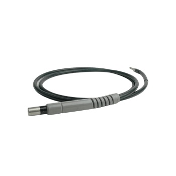
 Disconnect the LLG from the X-Cite and microscope adapter.
- Hold one end towards a bright window or overhead room light – DO NOT use an X-Cite or any other focused light source for this test!
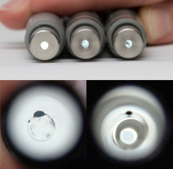
- Look at the quartz at the other end of the LLG.
- Bubble-free: quartz end will appear as a bright, solid circle; you may also be able to see a thin circular outline at the quartz/liquid interface.
- Bubbles at/near the quartz end: appear as dark spots, as small as 0.5mm in diameter or even as larger more defined spheres.
- Bubbles in the middle of the light guide: may not be well-defined spots, but will appear as dark shadows.
- In extreme cases, where the bubble is blocking the entire diameter of the light guide, no light will come through, even when pointing the distal end at a light source.
NOTE:
Liquid light guides universally are exposed to ultra-violet light which causes slow degradation in transmission properties. It is always a good idea to check your light guide if you notice a lower signal than normal (provided you have appropriate control slides). For more details on this and other helpful tips, download the X-Cite Maintenance Guide below.