Expertise in scientific imaging

NANOLIVE 3D Cell Explorer-fluo

COMBINE THE BEST OF TWO WORLDS

COMPLETE 3D SOLUTION

Combine high quality tomographic data with fluorescent markers

MULTIPLEXING

Explore up to 10 markers in parallel

EXTENDED LIVE CELL IMAGING

Limit cell damages caused by fluorescent markers, bleaching and phototoxicity

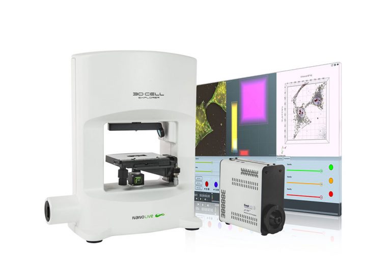
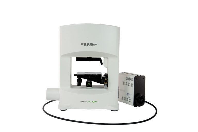
Nanolive Swiss High Precision

At Nanolive, we focus on innovative product development at the highest standards to deliver a high precision and quality research device for our international customers.

Available Configurations

DAPI configuration: DAPI + FitC + TritC

Live Cell Imaging configuration: FitC + TritC + Cy5

All-in-one configuration*: DAPI FitC + TritC Cy5

*TritC and Cy5 excitation filters are exchangeable on channel 3

The most powerful solution to image live cells

Multimodal Complete Solution for 3D Live Cell Exploration

Combine 3D refractive index analysis with a fully integrated 3 channel fluorescence module to image your live cells as they are and as long as you want. Put chemical information into structural context for new biological insights.

2D section of RI map
Overlay
Multi-channel epifluorescence
3D reconstruction based on RI map

Multiplexing

Transform 2D fluorescence into 3D cell tomography: Identify cell organelles through fluorescence and monitor non-invasively their structures and dynamics in 3D & marker-free. Explore fluorescence (3 channels) and Digital Stains (8 channels) simultaneously.

Co-localization for mitochondria
Co-localization for lysosomes
Co-localization for nuclear membrane
Co-localization for lipid droplets
Co-localization for plasma membrane
Co-localization for nucleus & nucleoli

Extended Live Cell Imaging

Image your live cells as long as you need. Limit cell damages caused by fluorescent markers, bleaching and phototoxicity.

Long-term imaging (11 hrs) of mouse pre-adipocytes. Mitochondria were labeled with mitTracker. A holotomographic image was taken every 15 seconds and a fluorescence image every 5 minutes.

Live FUCCI mESC imaged for over 48 hours

The video shows live FUCCI mESC that were imaged for over 48 hours with Nanolive’s 3D Cell Explorer-fluo. A holotomographic image was taken every 14 seconds while a double channel (green and red) epifluorescence image was only taken every 30 holotomographic frames (7:30 minutes). The difference in acquisition rate was performed to reduce fluorescence related phototoxicity. 

*Please note that the latest fluorescence image is displayed over the following 30 holotomographic images until a new one is acquired, for visualization purpose. The mismatch between the two signals is due to this image processing choice.

Discover More

Combine high quality RI tomographic data and fluorescence markers

You want to extend your understanding of cell structures and cell mechanisms? Thanks to our 3D tomographic technology you can now have a powerful platform that combines RI information with fluorescence markers.

Perform infinite live cell imaging

Limit cell damages caused by fluorescence markers, bleaching, and phototoxicity. The 3D Cell Explorer-fluo allows you to transform your chemical markers into physical ones which can be monitored for a limitless amount of time. 

Go beyond fluorescence limitation

You are frustrated by the limitations of fluorescence markers? You would like to go further? Combine them with Nanolive’s digital staining and push the limits forward.

Achieve specific cell analysis while preserving cell life

Forget about fixation! With the 3D Cell Explorer-fluo you can now observe your cells as they are: alive, 3D and in motion!

Technical Features 3D Cell Explorer-ƒluo